

Abstract
During pregnancy, iron is a primary requisite micronutrient for the developing fetal-placental unit and increased maternal erythrocyte mass expansion. Iron deficiency anemia in pregnancy (IDAP) remains a constant public health problem in our country where all of them receive routine iron supplementation.The mechanism involved in iron regulatory pathway across the placenta is more complex and less understood. Here we examined the hematological and biochemical parameters of mother and fetus, placental mRNA and protein expression of iron trafficking proteins in iron replete and deplete pregnant women.
Subjects who presented to the department of community health for child birth were screened and consenting patients (age<35 years and a gestational age≥36weeks) were included in the study. Maternal and cord blood samples were collected and a cut placental tissue (~0.5cm) was obtained at the time of delivery. Hematological and biochemical parameters were analysed according to standard methods. Serum ferritin was measured using chemiluminescent immunoassay. Maternal and cord blood serum hepcidin and GDF15 levels were measured using ELISA (DRG Diagnostics and Ray Biotech, respectively). Placental tissue RNA extracted using PARIS kit (Ambion) and cDNA was synthesised using RT2 first strand Kit (QIAGEN). The relative quantification of twenty two genes involved in iron uptake, export, transport and regulation was done using Custom RT2 PCR Array (SA Biosciences, QIAGEN). Western blot analysis was performed for significantly expressed genes. Statistical analysis was performed using SPSS software.
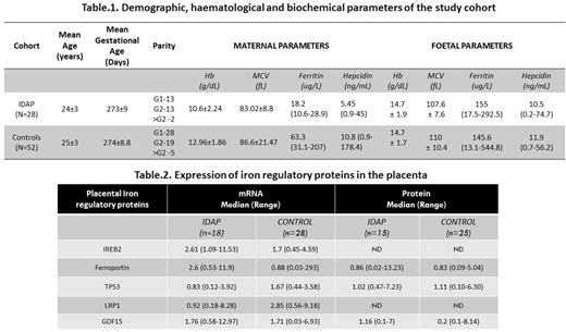
A total of one hundred and thirty eight pregnant women (n=138) were included in the study; 14 subjects were excluded due to unavailability of either maternal or cord blood serum samples. A cut off of ferritin<30ng/mL was taken to define iron deficiency since all of them were on regular iron supplementation during pregnancy. Of the 124 subjects, 28 subjects had iron deficiency anaemia (Hb<10.5g/dL and Ferritin<30ng/mL) and 52 were iron replete (Hb>10.5g/dL and Ferritin>30ng/mL). Forty four women who did not fulfil either of the criteria were not included for analysis. The demographic, hematological and biochemical parameters of the groups are tabulated (Table.1).
In the IDAP, even though the maternal serum ferritin levels were reduced, their fetal ferritin levels did not differ significantly from the control fetuses [IDAP fetuses- 139.8ng/mL, 15.4-300.5 vs Control fetuses - 143.4ng/mL, 13.1-461; p= 0.72]. No significant association was observed between ferritin and hepcidin levels either in the maternal or fetal serum. Interestingly, we found a negative correlation between maternal serum GD15 levels and fetal hepcidin: ferritin ratio (r=-0.439; p=0.025). Placental gene expression and protein expression was further analyzed (Table.2). Significantly higher gene expression of placental IREB2 [p=0.011], and ferroportin [p=0.000] were observed in iron deficient cohort. TP53 and LRP1 mRNA expressions were significantly lower in IDAP [p=0.002 & p=0.000]. Maternal serum ferritin had a significant positive correlation with placental expression of IREB2 (r=0.519; p=0.027) and LRP1 (r=0.470; p=0.049). At the protein level, placental GDF15 protein was significantly elevated in IDAP [p=0.04]. Placental GDF15 and ferroportin (FPN) was positively correlated with fetal ferritin (r=0.626; p=0.017 & r=0.552; p=0.041).
Even when the maternal iron stores are low, the fetus gets the priority according to the hierarchy of iron usage. In IDAP, we observed a higher expression of placental ferroportin and transferrin receptor which probably leads to the increased efflux of iron towards fetus. Elevated placental GDF15 and ferroportin may imply that they play a major role in accomplishing iron trafficking and has to be further explored.
No relevant conflicts of interest to declare.

This feature is available to Subscribers Only
Sign In or Create an Account Close Modal